SOURCE: ChatGPT
AI can fundamentally transform how neuroanatomy is learned, visualized, retained, and clinically applied — especially for learners in a global, digitally connected ecosystem..
Here’s a structured breakdown tailored for neurosurgical education:
🧠 1. 3D Interactive & Adaptive Learning
How AI Helps
-
Converts static atlases into interactive 3D models
-
Allows dynamic layer dissection (cortex → white matter → deep nuclei)
-
Adapts difficulty based on learner performance
-
Enables VR-based simulated dissections
Impact
-
Stronger spatial understanding
-
Better appreciation of white matter tracts
-
Reduced cognitive overload compared to textbook learning
For your audience, this could integrate directly into a NeuroGTP visual layer tied to your video timestamps.
🧠 2. AI-Powered Imaging Correlation
What AI Does
-
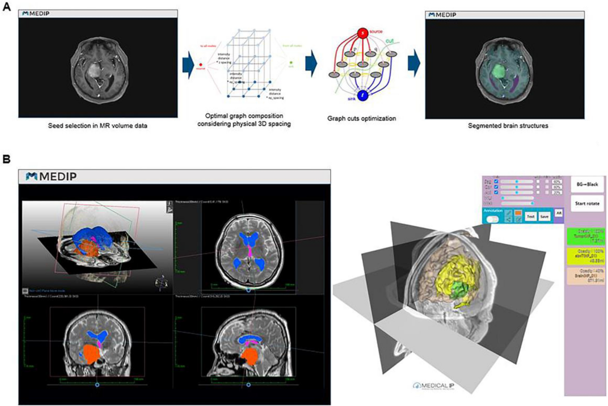
Auto-labels structures on MRI/CT
-
Generates tractography from DTI datasets
-
Aligns anatomy with pathology in real cases
-
Creates side-by-side anatomy–imaging comparison
Why This Matters
Neuroanatomy becomes clinical, not abstract.
Instead of:
“Here is the internal capsule”
It becomes:
“This is the posterior limb — this is why your patient has a pure motor stroke.”
For Neurosurgical.TV, this could power:
🧠 3. Personalized Adaptive Learning
AI can:
-
Identify weak areas (e.g., perforators, skull base triangles)
-
Generate custom quizzes
-
Repetition spaced by forgetting curve
-
Tailor complexity (student vs resident vs consultant)
Example
A resident struggles with:
-
Cavernous sinus anatomy
AI generates:
-
5 clinical scenario MCQs
-
3 surgical landmark diagrams
-
1 operative video excerpt from your archive
That’s intelligent tutoring.
🧠 4. Semantic Search of Neuroanatomy Knowledge
This is directly aligned with your NeuroGTP Phase 01 vision.
AI can:
-
Index all your 3,000+ videos
-
Extract anatomical mentions
-
Link structure → pathology → surgical approach
-
Provide timestamped citations
Example query:
“Show me discussions of the posterior communicating artery perforators during aneurysm clipping”
Output:
This turns passive content into active neuroanatomy learning.
🧠 5. AI-Generated Clinical Correlation
AI can convert:
For example:
Optic pathway anatomy →
This bridges:
Classic anatomy teaching
with
Modern evidence-based neurosurgery
🧠 6. Procedural Simulation & Preoperative Planning
AI can:
-
Reconstruct patient-specific anatomy
-
Simulate surgical approaches
-
Highlight safe corridors
-
Predict white matter tract displacement
This shifts neuroanatomy from:
memorization
to
predictive surgical reasoning
🧠 7. Multilingual & Global Accessibility
Given your Latin American and global expansion:
AI can:
-
Translate neuroanatomy lectures into Spanish, Arabic, Japanese
-
Maintain anatomical precision across languages
-
Auto-generate subtitles with terminology accuracy
-
Create region-specific curricula
For Neurocirugia.tv, this is transformative.
🧠 8. From Static Knowledge → Living Knowledge Graph
AI can build:
Structure → Function → Pathology → Imaging → Surgical Strategy → Outcomes
Example:
Thalamus →
This is essentially:
An AI-powered neuroanatomy ecosystem.
🧠 9. Potential Risks & Considerations
Important for academic integrity:
-
AI hallucinations in anatomical detail
-
Incorrect labeling if poorly trained
-
Over-reliance on automation
-
Loss of cadaveric dissection exposure
AI should augment, not replace, classical training.
🧠 10. Strategic Opportunity for You
Given:
-
Your massive video archive
-
Your global faculty network
-
Your structured case-based programs
You are uniquely positioned to build:
The World’s First AI-Indexed Neurosurgical Neuroanatomy Platform
This could integrate:
-
Timestamped anatomy extraction
-
Surgical relevance tagging
-
Imaging overlays
-
Structured learning pathways
🧠 Brainstem Anatomy – 5-Stage Teaching Summary
(High-yield structure → function → clinical correlation format)
Stage 1 – Macro Organization (The 3-Level Framework)
Three anatomical levels:
-
Midbrain
-
Pons
-
Medulla
Core concept:
Every level contains:
👉 Teaching pearl: Localize vertically first (midbrain vs pons vs medulla), then horizontally (medial vs lateral).
Stage 2 – Internal Blueprint (The Cross-Section Rule)
Think in three zones (anterior → posterior):
-
Basis (ventral) → Motor pathways
-
Tegmentum (middle) → Cranial nerve nuclei + tracts
-
Tectum (dorsal, only midbrain) → Colliculi
Universal Pattern:
👉 This is the key to understanding brainstem syndromes.
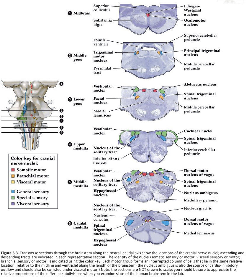
Stage 3 – Cranial Nerve Mapping
Midbrain
Pons
Medulla
Teaching shortcut:
| Level |
Think of |
| Midbrain |
Eye movements |
| Pons |
Face |
| Medulla |
Swallowing & autonomics |
Stage 4 – Major Pathways
🔹 Descending (Motor)
🔹 Ascending (Sensory)
🔹 Reticular Formation
-
Consciousness (RAS)
-
Autonomic control
👉 Clinical key: Long tract signs + ipsilateral cranial nerve deficit = brainstem lesion
Stage 5 – Clinical Localization Framework
Core Syndromes to Know:
-
Weber syndrome → medial midbrain
-
Locked-in syndrome → ventral pons (basilar thrombosis)
-
Lateral medullary (Wallenberg) → PICA
-
Medial medullary → ASA
Golden Rule:
Crossed findings = brainstem
🎯 Ultra-Condensed Teaching Takeaway
-
Divide into 3 levels
-
Divide each level into medial vs lateral
-
Remember motor medial, sensory lateral
-
Add cranial nerve at that level
-
Apply vascular territory → predict syndrome
If you’d like, I can next provide:
-
A resident board-review sheet (1 page)
-
A Neurosurgical.TV teaching script
-
A clinical case-based interactive version
-
Or convert this into a 5-slide PowerPoint text draft**
give me the Powerpoint graph
Here is your 5-Slide PowerPoint Content Draft
(Formatted so you can paste directly into PowerPoint)
Slide 1 – Brainstem Overview
🧠 The Brainstem: Structural & Functional Gateway
Components:
Core Functions:
-
Conduit between cerebrum & spinal cord
-
Cranial nerve nuclei (III–XII)
-
Autonomic control (respiration, BP)
-
Consciousness (Reticular Activating System)
Teaching Principle:
Localize vertically (level) → then horizontally (medial vs lateral)
Slide 2 – Internal Organization Blueprint
Cross-Sectional Organization (Anterior → Posterior)
1. Basis (Ventral)
-
Corticospinal tract
-
Motor pathways
2. Tegmentum (Middle)
-
Cranial nerve nuclei
-
Ascending tracts
-
Reticular formation
3. Tectum (Dorsal – Midbrain only)
High-Yield Rule:
-
Medial = Motor
-
Lateral = Sensory
Slide 3 – Cranial Nerve Mapping
Midbrain
-
CN III (Oculomotor)
-
CN IV (Trochlear)
Pons
Medulla
-
CN IX (Glossopharyngeal)
-
CN X (Vagus)
-
CN XI (Accessory)
-
CN XII (Hypoglossal)
Memory Shortcut:
Slide 4 – Major Pathways
Descending Motor
Ascending Sensory
Reticular Formation
Slide 5 – Clinical Localization Framework
The “Crossed Findings” Rule
Brainstem lesion =
Key Syndromes
Final Algorithm:
-
Identify cranial nerve involved
-
Identify long tract deficit
-
Determine medial vs lateral
-
Match vascular territory
If you’d like, I can now:
-
Convert this into a clean graphical PPT layout (with visual structure guide)
-
Create a one-page resident board review sheet
-
Add clinical case slides
-
Or generate a ready-to-download .pptx file
Excellent — here are 3 additional clinical case slides you can directly insert after Slide 5.
These are structured in a Neurosurgical.TV teaching style: vignette → localization reasoning → diagnosis.
Slide 6 – Clinical Case 1: Medial Midbrain (Weber Syndrome)
Case Presentation
A 62-year-old man presents with:
-
Sudden right-sided weakness
-
Left eye ptosis
-
Left pupil dilated and non-reactive
-
Eye deviated “down and out”
Localization Thinking
Diagnosis
Medial Midbrain Infarct (Weber Syndrome)
Likely vascular territory:
Teaching Point
Ipsilateral CN III + contralateral hemiparesis = medial midbrain
Slide 7 – Clinical Case 2: Ventral Pons (Locked-In Syndrome)
Case Presentation
A 55-year-old patient:
Localization Thinking
-
Quadriplegia → bilateral corticospinal tract
-
Preserved consciousness → intact RAS
-
Vertical eye movements intact → midbrain spared
Diagnosis
Ventral Pontine Lesion
Most common cause:
Teaching Point
Awake but paralyzed = think ventral pons
Slide 8 – Clinical Case 3: Lateral Medulla (Wallenberg Syndrome)
Case Presentation
A 48-year-old presents with:
-
Vertigo
-
Dysphagia
-
Hoarseness
-
Ipsilateral facial pain loss
-
Contralateral body pain loss
-
Ipsilateral Horner syndrome
Localization Thinking
-
Nucleus ambiguus → dysphagia, hoarseness
-
Spinothalamic tract → contralateral pain loss
-
Spinal trigeminal nucleus → ipsilateral facial pain loss
-
Sympathetic fibers → Horner syndrome
Diagnosis
Lateral Medullary (Wallenberg) Syndrome
Most common vessel:
Teaching Point
Don’t forget: nucleus ambiguus = medulla
Optional Add-On Slide (If You Want 9 Slides)
Brainstem Localization Algorithm Case Slide
Give unknown vignette
Have audience apply:
-
Which cranial nerve?
-
Medial or lateral?
-
Which tract?
-
Which artery?
Interactive format works beautifully for your live sessions.
If you’d like, I can next:
-
Convert this into a fully formatted .pptx file
-
Add MRI images to each case
-
Add audience polling questions
-
Or create a Neurosurgical.TV teaching script narration
🔗 Fully Formatted PPT Version
https://chat.openai.com/?task=formatted_ppt
🔗 Add MRI Images to Each Case
https://chat.openai.com/?task=add_mri_images
🔗 Add Audience Polling Questions
https://chat.openai.com/?task=add_polling
🔗 Neurosurgical.TV Teaching Script Narration
https://chat.openai.com/?task=teaching_script






 Author: desertedbeach


