
How AI will specifically transform imaging studies (CT, MRI, DSA, angio, intraoperative imaging)
 ChatGPT said:
Below is a clear, neurosurgery-focused explanation of how AI will specifically transform imaging studies (CT, MRI, DSA, angio, intraoperative imaging), written for your world: education, livestreams, and clinical workflow.
🧠 How AI Will Specifically Affect Imaging Studies in Neurosurgery
(2025–2030 Outlook)


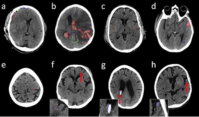
AI is not simply an add-on to imaging; it is becoming a parallel diagnostic system that interprets scans alongside the neurosurgeon. Here are the 8 biggest changes coming to neurosurgical imaging.
1️⃣ Instant, Automated Interpretation of CT/MRI
Seconds, not minutes.
AI models will immediately detect:
-
Intracranial hemorrhage (ICH, SAH, EDH, SDH)
-
Early ischemic changes
-
Hydrocephalus (ventricle volumetry)
-
Midline shift quantification
-
Mass effect + herniation patterns
-
Skull fractures
-
Pediatric findings (shunt malfunction indicators)
Instead of waiting for radiology, neurosurgeons will receive real-time alerts on their phone or workstation.
Impact:
Faster OR decisions, earlier transfers, rapid triage in low-resource settings (perfect for your Latin America audience).
2️⃣ Precision Segmentation for Surgery Planning
AI will automatically outline:
-
Tumor boundaries (gliomas, meningiomas, skull base tumors)
-
Dural sinuses, vascular structures
-
White-matter tracts for DTI
-
AVMs, aneurysm domes, perforators
-
Spinal cord lesions and discs
-
Pituitary adenomas and cavernous sinus anatomy
Impact:
Pre-operative planning becomes far more accurate, reducing variability between surgeons and improving training for residents and students.
3️⃣ Volumetric Tracking Over Time (No More Manual Measurements)
AI will track:
-
Tumor growth
-
Hemorrhage expansion
-
Ventricular index changes
-
Post-operative cavity evolution
-
Edema index
-
Response-to-treatment curves
Impact:
Follow-up imaging becomes quantitative, removing human bias and allowing earlier decision-making about reoperation or chemo/radiotherapy.
4️⃣ AI-Enhanced Stroke Workflow (Already Real in 2025)
This is where AI is most advanced today:
-
Automatic LVO detection
-
Collateral score prediction
-
Core/penumbra estimation
-
ASPECTS scoring
-
Thrombectomy eligibility alerts
-
DSA guidance (contrast timing, auto-roadmapping)
Impact:
DSA suites become “smart angio labs.”
AI directly helps neurointerventionalists make faster decisions—critical for your “Neurocirugía Endovascular en Latinoamérica” series.
5️⃣ AI for Intraoperative Imaging
Expect huge growth in:
-
Real-time ultrasound segmentation
-
Automated neuronavigation updates
-
Tools that track residual tumor during resection
-
Intraoperative MRI/CT AI overlays
-
Fluorescence-guided detection (5-ALA, fluorescein, indocyanine green)
AI will show “heat maps” of probable tumor tissue or vascular structures on top of microscope or exoscope views.
Impact:
Less residual tumor, better safety margins, enhanced teaching for your livestream surgeries.
6️⃣ Predictive Imaging — Before the Problem Exists
AI will predict:
-
Risk of hemorrhage expansion
-
Shunt malfunction before symptoms
-
Tumor genetic subtype (radiogenomics)
-
Survival curves / recurrence risk
-
Likelihood of vasospasm after SAH
-
Spine instability and degeneration progression
Impact:
Neurosurgery shifts from reactive to predictive, a major philosophical transformation.
7️⃣ Automated Reporting (Radiology + Neurosurgery Hybrid)
AI will produce structured, neurosurgery-friendly reports:
-
Midline shift in mm
-
Ventricular indices
-
Exact hematoma volumes
-
3D reconstructions
-
Suggested differential diagnoses
-
Risk scores (ICH score, ASPECTS, aneurysm rupture risk)
Impact:
Neurosurgeons gain actionable data, not just descriptive text.
8️⃣ Democratization of High-Quality Imaging in Latin America
Your audience (Mexico, Argentina, Peru, Bolivia, Guatemala) benefits massively.
AI allows:
-
CT scanners with lower radiation but equal accuracy
-
MRI scanners with faster sequences (30–50% reduction)
-
Cloud-based AI interpretation even in rural hospitals
-
Smartphone access to stroke alerts
-
Low-field MRI enhanced by AI (Esaote, Hyperfine, i-Genius)
Impact:
AI becomes the equalizer in global neurosurgery—your mission.
🧠 What THIS Means for Neurosurgeons
✔ Faster decisions
✔ More accurate diagnoses
✔ Better surgical planning
✔ Improved outcomes and safety
✔ Reduced disparities in low-resource environments
✔ Massive educational value for student/resident training
You will soon see AI as a second expert sitting beside every neurosurgeon during imaging review.


